Hypoxic-ischaemic encephalopathy (HIE) is the syndrome of disturbed neurological function in the term or late-preterm newborn that follows a peripartum interruption of cerebral oxygen delivery. It affects roughly 1–2 per 1,000 live births, is the commonest cause of neonatal encephalopathy in well-resourced settings, and is the only acquired newborn condition for which a neuroprotective therapy — therapeutic hypothermia — reduces death and major neurodevelopmental disability.
The ventilator is inseparable from that therapy. Hypothermia lowers metabolic rate and carbon dioxide production by roughly 6–7% per °C, so ventilator settings appropriate at 37.0 °C become over-ventilation at 33.5 °C and drive down the cerebral perfusion the injured brain most needs. Asphyxiated infants also frequently have pulmonary hypertension, myocardial dysfunction and acute kidney injury, each of which alters how gas exchange should be targeted. This chapter covers definition and staging, pathophysiology, the cooling protocol, and the ventilatory and neuromonitoring strategy that accompanies it.
Definition, Perinatal Asphyxia and Clinical Staging
From Asphyxia to Encephalopathy
Perinatal asphyxia is an interruption of placental gas exchange producing fetal hypoxaemia, hypercapnia and metabolic acidaemia. It is a physiological event; HIE is its clinical consequence, and the two are not interchangeable. Many acidotic infants are neurologically normal, and a substantial minority of neonatal encephalopathy is non-asphyxial — sepsis, stroke, metabolic disease or inborn errors of metabolism. The practical construct is therefore neonatal encephalopathy due to suspected HIE, in which biochemical evidence of intrapartum hypoxia coexists with a characteristic encephalopathy and alternatives have been excluded. Markers of an intrapartum event include an umbilical arterial pH of 7.0 or below, a base deficit of 16 mmol·L⁻¹ or greater, a lactate of 6 mmol·L⁻¹ or greater, an Apgar score of 5 or less at 10 minutes, or prolonged resuscitation; these confirm hypoxic exposure, not causation.
HIE is a clinically defined encephalopathy in the term or late-preterm newborn, attributable to a peripartum hypoxic-ischaemic event, characterised by altered consciousness, abnormal tone and reflexes, and seizures. A single episode of asphyxia may be the cause, but the diagnosis rests on the clinical syndrome, not on the cord gas alone.
The Sarnat Stages
Clinical staging follows Sarnat and Sarnat (1976), grading consciousness, neuromuscular control, primitive reflexes, autonomic function, seizure activity and EEG background. It underpins cooling eligibility and maps reasonably onto the deep grey matter injury that dominates outcome.
| Sarnat stage | Consciousness | Tone and posture | Primitive reflexes | Autonomic function | Seizures | EEG background | Course |
|---|---|---|---|---|---|---|---|
| I — mild | Hyperalert, irritable | Mild distal hypotonia, brisk reflexes | Strong suck and Moro | Sympathetic: mydriasis, tachycardia, tachypnoea | Absent | Normal or mildly abnormal | Recovery in 24–48 h; normal outcome |
| II — moderate | Lethargic, obtunded | Moderate hypotonia, proximal weakness | Weak suck, incomplete Moro, absent oculovestibular response | Parasympathetic: miosis, bradycardia, irregular respiration | Common (focal or multifocal) | Discontinuous, low-voltage or burst-suppression | Days to weeks; 25–40% death or disability |
| III — severe | Stupor or coma | Flaccid, absent reflexes | Absent suck, Moro and oculovestibular responses | Impaired: fixed pupils, apnoea, variable heart rate | Uncommon; decerebrate posturing | Isoelectric or persistent burst-suppression | High mortality; most survivors severely disabled |
The trajectory matters as much as the stage at presentation: progression from stage II to III, or failure of the aEEG background to recover, is ominous. Inter-observer agreement on clinical staging is only moderate, which is why examination is combined with amplitude-integrated electroencephalography (aEEG) and biochemical markers rather than used alone.
Hypothermia is indicated for moderate-to-severe encephalopathy (Sarnat II or III) or for seizures with an abnormal aEEG. Mild encephalopathy (Sarnat I) is generally excluded, because the risks of cooling outweigh any demonstrable benefit where the untreated prognosis is already good.
Pathophysiology of Hypoxic-Ischaemic Brain Injury
The defining concept of HIE is that the injury is not finished when the insult ends. Cerebral damage evolves through three overlapping phases, and the interval between the first and the second is the therapeutic window in which cooling works.
Primary Energy Failure
Reduced cerebral oxygen delivery causes oxidative phosphorylation to fail within minutes. Adenosine triphosphate (ATP) falls, the sodium–potassium ATPase can no longer maintain ionic gradients, and cells depolarise. Anaerobic glycolysis floods the tissue with lactate, and the resulting intracellular acidosis and calcium overload damage membranes and mitochondria. Accompanying hypercapnia and acidosis impair cerebral autoregulation, so perfusion becomes pressure-passive. This phase produces the acute cytotoxic oedema visible on early imaging.
The Latent Phase and the Therapeutic Window
After resuscitation, cerebral oxidative metabolism partially recovers and the infant may appear deceptively stable. This latent phase lasts roughly 1–6 hours: the electroencephalogram is typically suppressed, oxidative stress and inflammation are already being primed, and mitochondria are accruing the injury that will later declare itself. Because this interval precedes the second wave of cell death, cooling must begin within it; benefit falls steeply when hypothermia starts more than 6 hours after birth, which is why the therapeutic window defines the eligibility clock.
The latent phase is the only realistic opportunity to interrupt the secondary cascade. Once it has passed, the machinery of excitotoxicity, oxidative stress and programmed cell death is largely established, and cooling can only limit, not prevent, the injury.
Secondary Energy Failure
Between about 6 and 72 hours a second and often larger wave of injury unfolds. Excitatory amino acids, principally glutamate, over-activate N-methyl-D-aspartate (NMDA) and AMPA receptors, permitting a lethal calcium influx. Mitochondrial permeability transition follows, with cytochrome c release and caspase activation. Reactive oxygen and nitrogen species overwhelm immature antioxidant defences, and activated microglia release pro-inflammatory cytokines that amplify the damage; cell death proceeds through mixed apoptosis, necrosis and necroptosis. Cooling attenuates this phase, which is also when seizures and haemodynamic instability cluster.
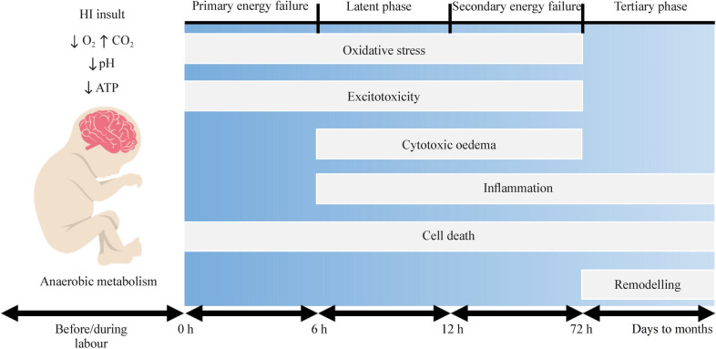 Figure 1: The evolution of hypoxic-ischaemic injury — reduced cerebral oxygen delivery, hypercapnia and falling pH and ATP during the insult, followed by the latent interval and the secondary wave that hypothermia is designed to interrupt. (Source: Babbo CC, Mellet J, van Rensburg J, et al. World J Pediatr 2024. DOI: 10.1007/s12519-024-00836-9).
Selective Vulnerability
Regional vulnerability is not uniform. Acute profound asphyxia — cord occlusion, placental abruption — preferentially injures the deep grey matter: the putamen, ventrolateral thalamus, hippocampus and perirolandic cortex, structures with the highest metabolic demand, densest NMDA receptor expression and terminal penetrating arterial supply. This predicts extrapyramidal cerebral palsy with relative cognitive sparing. Prolonged partial asphyxia instead injures the parasagittal watershed cortex and subcortical white matter, producing spastic quadriparesis; in preterm infants the vulnerable population shifts to the periventricular white matter. Loss of autoregulation compounds the risk, because perfusion depends on the pressure head:
$$CPP = MAP - ICP$$where $CPP$ = cerebral perfusion pressure ($\text{mmHg}$); $MAP$ = mean arterial pressure ($\text{mmHg}$); and $ICP$ = intracranial pressure ($\text{mmHg}$). In neonates $ICP$ is low and difficult to measure, so $MAP$ is the practical surrogate and systemic hypotension is directly injurious.
Therapeutic Hypothermia: Evidence, Eligibility and Protocol
The Evidence Base
CoolCap (2005), the NICHD whole-body trial (2005) and TOBY (2009), together with a Cochrane systematic review, established cooling as standard care. Pooled data show reduced death or major neurodevelopmental disability at 18–24 months, with a relative risk of roughly 0.75 and a number needed to treat of about 7–9. Benefit is greatest when cooling starts early and the encephalopathy is moderate rather than severe. Notably, the HELIX trial in low-resource settings without equivalent intensive monitoring found increased mortality — cooling is a package, not a temperature.
Eligibility and the Therapeutic Window
Eligibility combines evidence of an intrapartum hypoxic event, an appropriate postnatal age, and moderate-to-severe encephalopathy.
| Domain | Typical requirement |
|---|---|
| Gestational age and weight | $\geq 36$ weeks and $\geq 1{,}800$–$2{,}000\ \text{g}$ |
| Postnatal age | Less than 6 hours at the start of cooling |
| Evidence of asphyxia | pH $\leq 7.0$, base deficit $\geq 16\ \text{mmol}\cdot\text{L}^{-1}$, lactate $\geq 6\ \text{mmol}\cdot\text{L}^{-1}$, Apgar $\leq 5$ at 10 min, or prolonged resuscitation |
| Neurological status | Sarnat stage II or III, or clinical seizures |
| Neurophysiology | Moderately or severely abnormal aEEG background, or electrographic seizures |
| Exclusions | Major congenital anomaly, uncontrolled bleeding, overwhelming multiorgan failure, or a setting without neonatal intensive care |
graph TD
    A["Term or late-preterm infant with suspected HIE"] --> B{"Evidence of intrapartum asphyxia?<br/>pH <= 7.0, base deficit >= 16 mmol/L,<br/>Apgar <= 5 at 10 min, or resuscitation"}
    B -->|"No"| C["Manage as neonatal encephalopathy;<br/>investigate alternative diagnoses"]
    B -->|"Yes"| D{"Age < 6 h AND GA >= 36 weeks<br/>AND weight >= 1800 g?"}
    D -->|"No"| E["Cooling not indicated;<br/>provide standard neuroprotective intensive care"]
    D -->|"Yes"| F{"Moderate-to-severe encephalopathy?<br/>Sarnat II-III, seizures, or<br/>moderately/severely abnormal aEEG"}
    F -->|"No"| E
    F -->|"Yes"| G{"Any exclusion present?<br/>major anomaly, uncontrolled bleeding,<br/>overwhelming multiorgan failure"}
    G -->|"Yes"| E
    G -->|"No"| H["Begin whole-body cooling:<br/>core target 33.5-34.5 C for 72 h"]
    H --> I["Ventilate to normocapnia:<br/>PaCO2 35-45 mmHg, avoid < 30 mmHg"]
    I --> J["Target SpO2 94-98%; avoid hyperoxia<br/>and wean FiO2 promptly"]
    J --> K{"PPHN suspected?<br/>labile hypoxaemia, pre-/post-ductal gradient"}
    K -->|"Yes"| L["Recruit lung, support systemic BP,<br/>consider iNO; never hyperventilate"]
    K -->|"No"| M["Lung-protective ventilation:<br/>Vt 4-6 mL/kg, PEEP 4-6 cmH2O"]
    L --> N["After 72 h rewarm 0.2-0.5 C per hour;<br/>recheck gases as CO2 production rises"]
    M --> N
The Cooling Protocol
Whole-body cooling is the commonest approach; selective head cooling with a cooling cap is an acceptable alternative. The target is a core temperature — measured oesophageally, rectally or nasopharyngeally — of 33.5–34.5 °C, maintained for 72 hours. Servo-controlled devices regulate the deviation from target:
$$\Delta T = T_{core} - T_{target}$$where $\Delta T$ = temperature deviation ($^\circ\text{C}$); $T_{core}$ = measured core temperature ($^\circ\text{C}$); and $T_{target}$ = the protocol midpoint of 34.0 °C ($^\circ\text{C}$), maintained within approximately $\pm 0.5\ ^\circ\text{C}$. Rewarming must be controlled: 0.2–0.5 °C per hour to a normothermic 36.5–37.0 °C over at least 6 hours, because rewarming raises cerebral metabolic demand and carbon dioxide production, may unmask seizures, and risks rebound pulmonary hypertension.
A core temperature above 38 °C during or after cooling is independently associated with worse outcome. Every rewarming phase must be paired with gas-exchange reassessment, because the minute ventilation that suited the hypothermic infant becomes inadequate as metabolic rate climbs.
When Cooling Is Not Indicated
Cooling should not be offered for mild encephalopathy (Sarnat I), for infants presenting after 6 hours of age, or for preterm infants below roughly 36 weeks, in whom the evidence is absent and harm — bradycardia, coagulopathy, death — is real. It is also inappropriate with major congenital or chromosomal anomalies, uncontrolled haemorrhage, overwhelming multiorgan failure, or in units without continuous temperature control, invasive monitoring and experienced intensive care. In these circumstances, meticulous normothermia, seizure control, ventilation to normocapnia and supportive care remain the standard.
The Interaction Between Cooling and Ventilation
Carbon Dioxide, Cerebral Blood Flow and Hypocapnia
Carbon dioxide is the most powerful physiological regulator of cerebral blood flow in the newborn. Over the physiological range, flow changes by roughly 3–4% per mmHg of PaCO2, so a fall from 40 to 30 mmHg reduces cerebral blood flow by about 30%. This reactivity can be approximated around normocapnia:
$$CBF \approx CBF_{0}\left[1 + k\left(PaCO_2 - 40\right)\right]$$where $CBF$ = cerebral blood flow ($\text{mL}\cdot100\ \text{g}^{-1}\cdot\text{min}^{-1}$); $CBF_{0}$ = cerebral blood flow at a $PaCO_2$ of 40 mmHg; $k$ = the CO2 reactivity coefficient (approximately 0.03–0.04 per mmHg in the term newborn); and $PaCO_2$ = arterial carbon dioxide tension ($\text{mmHg}$). Hypocapnia therefore vasoconstricts the cerebral circulation at the moment the injured brain is most vulnerable to ischaemia, and it shifts the oxyhaemoglobin dissociation curve left so that less oxygen is unloaded to tissue. Because cooling lowers carbon dioxide production, the trap is entirely predictable: unless minute ventilation falls with temperature, the infant becomes hypocapnic.
A PaCO2 below 30 mmHg is associated with worse neurodevelopmental outcome in HIE. Hyperventilation is never an acceptable treatment for pulmonary hypertension in this population; target normocapnia and treat the pulmonary vasculature pharmacologically.
Oxygen, Hyperoxia and Pulmonary Vascular Tone
Hyperoxia is an equally avoidable insult, because reoxygenation of reperfused tissue generates reactive oxygen species and an arterial oxygen tension above about 100 mmHg increases oxidative injury without improving outcome. Saturation, not tension, carries the bulk of the oxygen:
$$CaO_2 = 1.34 \times Hb \times SaO_2 + 0.003 \times PaO_2$$where $CaO_2$ = arterial oxygen content ($\text{mL}\ \text{O}_2\cdot\text{dL}^{-1}$); $Hb$ = haemoglobin concentration ($\text{g}\cdot\text{dL}^{-1}$); $SaO_2$ = arterial oxygen saturation (fraction); and $PaO_2$ = arterial oxygen tension ($\text{mmHg}$). Above an $SaO_2$ of 95%, further rises in $PaO_2$ add negligible oxygen content but substantial oxidative risk.
Pulmonary Hypertension and Persistent Fetal Circulation
Asphyxia and hypothermia interact to raise pulmonary vascular resistance, and pulmonary hypertension with persistent fetal circulation complicates a significant minority of cooled infants. Hypothermia itself increases pulmonary vascular resistance, bradycardia is expected, and asphyxial myocardial dysfunction reduces the ability to tolerate a pressure-loaded right ventricle. Management follows the principles of any pulmonary hypertension — recruit the lung without overdistending it, defend systemic blood pressure, and consider inhaled nitric oxide — with the added constraint that hyperventilation is forbidden. Labile hypoxaemia or a pre- to post-ductal saturation gradient should prompt echocardiography, not a change in ventilator settings.
Ventilatory Management of the Asphyxiated Newborn
Establish effective ventilation, secure normocapnia, correct hypoxia and hypotension, treat seizures and hypoglycaemia, and start cooling within 6 hours. Each is time-critical, and none can be deferred for the convenience of the next.
Lung-Protective Ventilation and Normocapnia
Most infants with moderate-to-severe HIE require intubation, usually for apnoea, poor respiratory drive, sedation during cooling, or coexisting meconium aspiration. Conventional pressure-limited or volume-targeted ventilation with a tidal volume of 4–6 mL·kg⁻¹ and PEEP of 4–6 cmH₂O limits volutrauma and atelectrauma while avoiding the hyperventilation that pressure-limited modes readily produce as compliance and metabolic rate fall. Target a PaCO2 of 35–45 mmHg, with the locally agreed range documented and audited.
Neonatal blood gases are reported at 37 °C and are managed uncorrected (alpha-stat), not temperature-corrected. Do not adjust ventilation for a calculated temperature-corrected PaCO2; doing so systematically over-ventilates the cooled infant.
Oxygen Targeting
Target a pre-ductal SpO2 of 94–98% and wean inspired oxygen promptly once that is achieved. Continuous pulse oximetry with alarm limits, and blood gases that include PaO2, allow hyperoxia to be detected and corrected; the goal is normoxaemia, not the highest saturation that can be achieved.
Sedation, Analgesia and Shivering
Cooling is distressing and provokes shivering, which generates heat, defeats the protocol and raises oxygen consumption. A continuous morphine or fentanyl infusion smooths cooling and improves ventilator tolerance, but hypothermia reduces hepatic clearance, so opioid accumulation and ileus are common. Titrate to effect and review daily rather than running a fixed rate for 72 hours.
Shivering both defeats cooling and raises oxygen consumption, so it must be treated. But hypothermia slows hepatic clearance: review the opioid infusion daily and expect ileus, urinary retention and delayed awakening.
Apnoea, Seizures and Neuromonitoring-Driven Care
Central apnoea is common and is an absolute indication for ventilatory support; it usually improves as the encephalopathy resolves. Seizures occur in a large proportion of cooled infants, are frequently electrographic without clinical correlate, and increase cerebral metabolic demand. Phenobarbital remains first-line, with levetiracetam or midazolam second-line, titrated against continuous or amplitude-integrated EEG rather than clinical signs alone.
Cardiovascular and Renal Consequences
Asphyxial myocardial dysfunction, together with the vasoconstriction and reduced cardiac output of cooling, makes hypotension likely; treat it with modest volume aliquots and inotropes or vasopressors — dopamine, dobutamine or adrenaline — guided by perfusion and echocardiography. Acute kidney injury is common, presenting as oliguria and a rising creatinine, and hyponatraemia from inappropriate antidiuretic hormone secretion is frequent. Monitor fluid balance closely, avoid nephrotoxic drugs, and keep glucose within the normal range, because both hypoglycaemia and hyperglycaemia worsen injury.
Neuromonitoring and Prognostication
Amplitude-Integrated EEG
Amplitude-integrated EEG (aEEG) is the workhorse of HIE monitoring. A single- or two-channel tracing obtained within the first 6 hours both supports cooling eligibility and provides early prognostic information: a continuous normal-voltage background predicts a favourable outcome, whereas burst-suppression, a low-voltage or flat trace — or failure of the background to recover over 24–48 hours — predicts death or severe disability. The aEEG also detects electrographic seizures that clinical examination misses, although its compressed display can miss brief or focal discharges, so positive findings should be confirmed with conventional EEG where available.
Cerebral Near-Infrared Spectroscopy
Cerebral near-infrared spectroscopy (NIRS) measures regional cerebral oxygen saturation and allows the fractional tissue oxygen extraction to be calculated. A falling cerebral saturation with a rising extraction fraction suggests inadequate perfusion or excessive cerebral oxygen demand, and the technique reveals the cerebral consequences of hypocapnia or hypotension in real time. NIRS is a trend monitor, however, not a validated stand-alone outcome predictor; it informs adjustments to ventilation, blood pressure and sedation rather than replacing aEEG or imaging.
Magnetic Resonance Imaging
Magnetic resonance imaging (MRI) is the reference standard for structural prognostication. Diffusion-weighted imaging detects the cytotoxic oedema of the deep grey matter with high sensitivity during the first days of life, but the signal can pseudonormalise around the end of the first week, so early scans may underestimate injury. The optimal window for prognostication is generally between about 8 and 14 days, when T1, T2 and diffusion sequences together define the pattern and extent of injury. Basal ganglia and thalamic injury carries the worst prognosis, a purely watershed pattern is intermediate, and a normal study predicts a good outcome; magnetic resonance spectroscopy adds prognostic value through the lactate-to-N-acetylaspartate ratio. Because no single modality is reliable in isolation, prognostication should be multimodal — serial examination, aEEG recovery, MRI and feeding — and communicated with appropriate humility.
Key Takeaways
- HIE is a clinical syndrome, not a biochemical diagnosis. Distinguish perinatal asphyxia from encephalopathy due to suspected HIE, and actively exclude non-asphyxial causes.
- Stage encephalopathy with the Sarnat system, supported by aEEG. Moderate-to-severe disease (Sarnat II–III) or seizures is the gateway to cooling; mild disease is generally not cooled.
- The injury evolves through primary energy failure, a latent phase and secondary energy failure. The latent phase is the therapeutic window, so cooling begun after 6 hours of age has little benefit.
- Cool eligible infants to a core temperature of 33.5–34.5 °C for 72 hours, then rewarm at 0.2–0.5 °C per hour with continuous gas-exchange reassessment.
- Hypothermia lowers metabolic rate and carbon dioxide production: unless minute ventilation is reduced, the infant becomes hypocapnic and cerebral perfusion falls. Target PaCO2 35–45 mmHg and never hyperventilate below 30 mmHg.
- Target SpO2 94–98% and avoid hyperoxia. Anticipate pulmonary hypertension with persistent fetal circulation, managing it by lung recruitment, systemic blood-pressure support and consideration of inhaled nitric oxide.
- Use lung-protective settings (Vt 4–6 mL·kg⁻¹, PEEP 4–6 cmH₂O), alpha-stat blood gas interpretation, adequate sedation for shivering, and seizure treatment titrated to EEG.
- Prognosticate multimodally with serial examination, aEEG background recovery and MRI at 8–14 days; deep grey matter injury predicts the worst outcome.